Management of supravalvular aortic stenosis with the Doty technique, in a South American pediatric center

Manejo de la estenosis aórtica supravalvular con la técnica Doty en un centro pediátrico sudamericano

Eliana B. Yagual-Gutiérrez1*, Isabel M. Freire-Barrezueta2, Mercedes C. Hernandez-Duarte3, Isabel J. Calero-Solis1, David F. Maldonado-González1, Darwin E. Zhune-Villagran1

1Department of Pediatric Cardiovascular Surgery; 2Department of Pediatric Cardiology; 3Pediatric Cardiovascular Intensive Care. Children Hospital Roberto Gilbert Elizalde, Guayaquil, Ecuador


*Correspondence: Eliana B. Yagual-Gutiérrez. E-mail: draelianayagual@gmail.com

Date of reception: 28-04-2024
Date of acceptance: 15-07-2024
DOI: 10.24875/ACM.24000089
Available online: 29-07-2024
Arch Cardiol Mex. 2025;95(2):215-219

Abstract

Supravalvular aortic stenosis (SVAS) is a congenital malformation associated with Williams-Beuren syndrome. It is characterized by severe stenosis of the sinotubular junction. The smooth muscle layer is thickened due to an alteration of the Elastin gene. The treatment is surgical and there are several techniques to relieve obstruction which range from the single-patch technique to the replacement of all three aortic sinuses with three patches. However, the optimal technique is still unclear. The objective of this article is to carry out a bibliographic review of surgical techniques for SVAS and the description of the application of the Doty technique for a 4-year-old infant at the Roberto Gilbert Elizalde Hospital.

Keywords: Doty technique; Williams-Beuren syndrome; Supravalvular aortic stenosis


Resumen

La estenosis aórtica supravalvular es una malformación congénita asociada al síndrome de Williams-Beuren. Se caracteriza por una severa estenosis en la unión sinotubular. La capa de músculo liso se engrosa debido a una alteración en el gen Elastina. El tratamiento es quirúrgico y existen varias técnicas para aliviar la obstrucción, que van desde la técnica de un solo parche hasta el reemplazo de los tres senos aórticos con tres parches. Sin embargo, la técnica óptima aún no está clara. El objetivo de este artículo es realizar una revisión bibliográfica de las técnicas quirúrgicas para estenosis valvular aórtica supravalvular y describir la aplicación de la técnica de Doty en una niña de 4 años en el Hospital Roberto Gilbert Elizalde.

Palabras clave: Técnica de Doty; Síndrome de Williams-Beuren; Estenosis aórtica supravalvular


Introduction

Supravalvular aortic stenosis (SVAS) is a rare congenital malformation, associated with Williams–Beuren syndrome (WBS). It is caused by the alteration of the elastin gene on chromosome 7q11.23. It is transmitted in a dominant manner, with the majority of cases being sporadic1. The transmission of the disease from an affected parent is rare1-3.

SVAS is characterized by severe luminal narrowing of the sinotubular junction (STJ) and the ascending aorta, it is less frequently characterized with the aortic arch and descending aorta4,5.

Several techniques have been developed to repair SVAS, the first technique was introduced in 1966 by McGoon, who performed a single patch augmentation of the non-coronary sinus6. Doty et al. described an extended aortoplasty of the non-coronary sinus and the right coronary sinus using a trouser patch in 19777. In 1988, Brom reported a technique for augmenting the three sinuses of Valsalva8.

To date, no surgical technique has demonstrated clear superiority. In our setting, as the national reference center for congenital heart disease, we aim to present a case of Doty surgery on a 4-year-old girl with symptomatic SVAS, along with a literature review on the current proposal for the surgical management of SVAS.

Description of the clinical case

The 4-year-old female child was referred to our hospital from a Ministry of Health Hospital at the age of three. She presented with delayed psychomotor development and irritability. She was diagnosed with WBS, Karyotype 46, XX. Physical examination revealed facial dysmorphia characterized by a broad forehead, short and wide nose, slight palpebral and epicanthal obliquity, a small jaw, and prominent lips. A systolic murmur of intensity IV/VI with a rhomboid characteristic (crescendo-decrescendo) and irradiation to the neck was observed in the aortic focus. The echocardiogram showed flow acceleration at the level of the aortic supravalvular junction.

Complementary studies included a chest X-ray that revealed excavation of the lung button (Fig. 1) with no other relevant findings. The electrocardiogram showed ST depression, left ventricular growth with a positive Sokolow-Lyon index, suggesting signs of systolic overload and myocardial ischemia. The two-dimensional transthoracic echocardiogram confirmed concentric left ventricular hypertrophy; normal mobility; preserved ejection fraction of 70%; tri-leaflet aortic valve without alterations; the presence of severe SVAS with a maximum gradient of 116 mmHg and a mean gradient of 65 mmHg. The aortic annulus measured 10 mm (z-score-1.7); the sinuses of the Valsalva measured 14mm (z-score –1.3); and STJ measured 8 mm (z-score –3.6) (Fig. 2). These findings were confirmed with chest angiotomography (Fig. 3). Based on these findings, the diagnosis of SVAS was confirmed, and the decision was made to perform Doty’s procedure to resolve the stenosis. The girl is currently asymptomatic with regular cardiovascular follow-ups every 6 months.

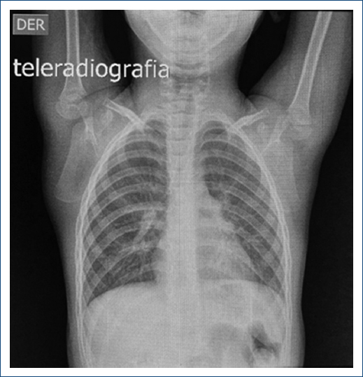
Figure 1. Posteroanterior chest radiograph with a normal cardiothoracic index and button excavation pulmonar.


Figure 2. Two-dimensional transthoracic echocardiogram, A: parasternal long axis shows supravalvular aortic stenosis (red arrow), B: Doppler echocardiogram shows flow acceleration at the level of the STJ (black arrow) of 5.40 m/s.


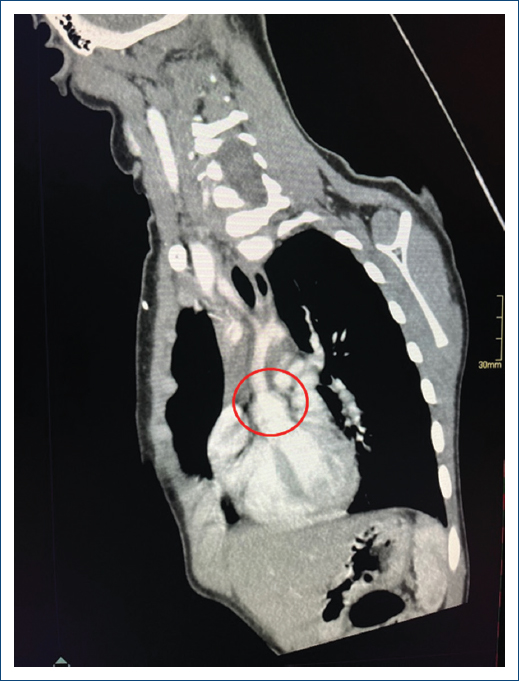
Figure 3. Chest angiotomography, orthogonal and long-axis plane of the aortic roof, demonstrating the typical hourglass image (red circle), with an annulus of 17 mm (z 0.38) and an STJ of 8.8mm (z-3.6).


Surgical technique

The surgery consisted of right atrial cannulation and aortic cannulation near the brachiocephalic trunk. Left cavity drainage was achieved through the right superior pulmonary vein, and the aorta was clamped. A single dose of Nido cardioplegia was administered anterogradely and directly into the coronary ostium. The aorta was sectioned at the point of maximum stenosis, addressing the non-coronary sinus to allow better exposure of the sinuses containing the coronary ostium. The non-coronary sinus was resected, and an incision was made 2-3 mm from the nadir of the sinus of Valsalva to the left of the ostium of the right coronary artery in the right coronary sinus. The left supracoronary crest was resected due to intimal hyperplasia.

A pant-shaped polytetrafluoroethylene patch was used to create the neo-sinuses of Valsalva and increase the diameter of the ascending aorta (Fig. 4). The aortic clamping time was 75 min, and the extracorporeal circulation time was 105 min, leading to a return to sinus rhythm. Transesophageal echocardiography performed during the surgery confirmed adequate aortic valve coaptation and the absence of a significant gradient at the level of the STJ.

Figure 4. Intraoperative image, A: aorta with hourglass image (thick lines). B: pantaloon-shaped patch, Doty technique.


In the immediate post-operative period, bleeding was greater than usual, necessitating re-intervention. The patient was discharged 15 days after surgery with cardiological follow-up and is awaiting a new control chest angiotomography.

Discussion

SVAS is a rare and complex disease often associated with multiple cardiac anomalies and syndromes, typically linked to WBS, of which around 69% are diagnosed with SVAS. It has an early mortality rate of 4.2% and a late mortality rate of 0.6%9,10.

Following the proposed guidelines by Mongé et al.4, the criteria for surgery include a significant gradient in the left ventricular outflow tract of 40-50 mmHg along with a significant narrowing of the STJ. Another criterion is a gradient of 30-40 mmHg at the level of the STJ plus significant left ventricular hypertrophy. Our patient met the latter criteria, with the maximum gradient being 116 mmHg at the level of the STJ.

Due to its rarity, there exists a limited cohort of patients, resulting in many different surgical techniques evolving from 1966 to the present day for SVAS correction, this diversity of approaches complicates the decision-making process regarding the selection of an ideal surgical technique11,12.

Stamm et al.11 conducted a study involving 76 patients who underwent surgery utilizing the three described techniques over a span of 41 years. In most patients, they included the Doty technique. They concluded that the insertion of at least one inverted bifurcated patch in the right coronary and non-coronary sinuses is not technically demanding. It will likely lead to improvements in long-term survival with lower reoperation rates.

In 2022, Ibarra et al.12 reported the follow-up of 89 patients, among whom 31 underwent surgery with the single patch technique and 58 with the Doty technique. With a mean age at surgery was 2.5 years and a median follow-up of 5.8 years, they observed a greater change in the STJ with suitable diameters in patients treated with the Doty technique, along with greater freedom of reoperation in this group.

Meccanici et al.9, being the first meta-analysis reported in 2024, included 23 publications with a total of 1,472 patients. The mean age at the time of surgical repair was 4.7 ± 5.8 years, with 49.6% having WBS, and a mean follow-up of 6.2 years.

The single-patch technique was reported in 43.6%, followed by the Doty technique in 33.2% of cases. The higher number of patients with the single-patch technique is due to its longer period of usage, but in different analyzed reports11-13, the Doty technique showed lower reoperation rates and higher survival.

For this reason, at our center, Doty Surgery is performed as the technique of choice for SVAS.

The early complications reported with the Doty technique11 include severe arrhythmias, acute myocardial ischemia, and the need for ECMO. In our case, the main complication was post-surgical bleeding, requiring re-exploration. The porosity of the artificial patch used is likely the cause of the post-surgical event.

The post-operative transthoracic echocardiogram showed no significant gradients at the level of the STJ, being < 5 mmHg, with a lesion-free aortic valve.

The late complications reported by Meccanici, showed microsimulation-based event risks in the first 30 post-operative years after congenital SVAS repair: the risk of myocardial infarction was 8.1% (95% Confidence interval [CI]: 7.3-9.9). %), and the risk of reintervention was 31.3% (95% CI: 29.6-33.4%).

To avoid one of the major complications of SVAS repair related to the coronary arteries, Lizhi et al.13 modified the Doty Technique. This was applied to 6 patients and compared to 64 patients who received the traditional Doty technique, yielding promising results. These require meticulous long-term follow-up to determine their place in the surgical armamentarium for SVAS.

Furthermore, the case we present is important because it involves a rare pathology and requires more publications to elucidate the ideal technique for SVAS. We have opted to use the surgical technique that has demonstrated better long-term survival. The successful application of the Doty technique in this specific case reinforces its role as an effective option in managing SVAS, especially considering the ease of performing the surgical technique, lower complication rates, and the low gradient at the STJ.

Conclusion

The reconstruction of the STJ and aorta is feasible with the Doty technique. However, it requires lifelong monitoring of the cardiovascular system to evaluate residual stenosis and coronary obstruction, utilizing serial echocardiograms and chest angiotomography.

Funding

None.

Conflicts of interest

None.

Ethical considerations

Protection of humans and animals. The authors declare that no experiments involving humans or animals were conducted for this research.

Confidentiality, informed consent, and ethical approval. The authors have followed their institution’s confidentiality protocols, obtained informed consent from patients, and received approval from the Ethics Committee. The SAGER guidelines were followed according to the nature of the study.

Declaration on the use of artificial intelligence. The authors declare that no generative artificial intelligence was used in the writing of this manuscript.

References

1. Williams JC, Barratt-Boyes BG, Lowe JB. Supravalvular aortic stenosis. Circulation. 1961;24:1311-8.

2. Beuren AJ, Schulze C, Eberle P, Harmjanz D, Apitz J. The syndrome of supravalvular aortic stenosis, peripheral pulmonary stenosis, mental retardation and similar facial appearance. Am J Cardiol. 1964;13:471-83.

3. Ruiz L, Lima MM, Quijada B, Aguirre U, Miguel A. Williams-Beuren syndrome:about a case. Rev Venez Endocrinol Metal. 2013;vol.11, n.1, pp.49-54.

4. MongéMC, Eltayeb OM, Costello JM, Jhonson JT, Popescu AR, Rigsby CK, et al. Brom aortoplasty for supravalvular aortic stenosis. World J Pediatr Congenit Heart Surg. 2018;9:139-46.

5. Hussein N, Honjo O, Barron DJ, Yoo SJ. Supravalvular aortic stenosis repair:surgical training of 2 repair techniques using 3D-printed models. Interact Cardiovasc Thorac Surg. 2021;33:966-8.

6. Rastelli GC, McGoon DC, Ongley PA, Mankin HT, Kirklin JW. Surgical treatment of supravalvular aortic stenosis. Report of 16 cases and review of literature. J Thorac Cardiovasc Surg. 1966;51:873-82.

7. Doty DB, Polansky DB, Jenson CB. Supravalvular aortic stenosis. Repair by extended aortoplasty. J Thorac Cardiovasc Surg. 1977;74:362-71.

8. Brom AG. Obstruction of the left ventricular outflow tract. In:Cardiac Surgery:Safeguards and Pitfalls in Operative Technique. Rockville, MD:Aspen Publishers;1988. 276-80.

9. Meccanici F, Notenboom ML, Meijssen J, Smit V, Van de Woestijne PC, Van den Bosch AE, et al. Long-term surgical outcomes of congenital supravalvular aortic stenosis:a systematic review, meta-analysis and microsimulation study. Eur J Cardiothorac Surg. 2024;65:ezad360.

10. Pober B, Johnson M, Urban Z. Mechanisms and treatment of cardio-vascular disease in Williams-Beuren syndrome. J Clin Invest. 2008;118:1606-15.

11. Stamm C, Kreutzer C, Zurakowski C, Nollert G, Friehs I, Mayer JE, et al. Forty-one years of surgical experience with congenital supravalvular aortic stenosis. J Thorac Cardiovasc Surg. 1999;118:874-85.

12. Ibarra C, Spigel Z, John R, Binsalamah ZM, Adachi I, Heinle JS, et al. Surgical techniques in management of supravalvular aortic stenosis in children. Ann Thorac Surg. 2021;111:2021-7.

13. Lizhi LV, Lang X, Zhang S, Wang C, Jin Y, Wang Q. Clinical results of a modified doty's technique for supravalvular aortic stenosis. J Clin Med. 2023;12:1731.